Showing posts with label diagnostic. Show all posts

Friday, July 12, 2013

British Laminitis Research: Tracking Normal Horses Who Later Develop Pasture Laminitis Vs Those Who Don't

Typical feet of a pony with pasture laminitis. Could laminitis be prevented by identifying likely-to-founder ponies early in their lives? A British research project aims to track normal ponies who, later in life, do and don't develop pasture-associated laminitis. (Nicola Menzies-Gow photo)

Dr Nicola Menzies-Gow of the Royal Veterinary College has been awarded a grant of £42,000 (approximately $65,000US) by Great Britain's Animal Welfare Foundation to work on a study: "Markers of equine laminitis predisposition: Searching for potential future diagnostic test". The award was announced this week by the college.

Thursday, April 14, 2011

Dr. David Hood Launches the Hoof Diagnostic and Rehabilitation Clinic in Texas

David M. Hood DVM, PhD will launch a new chapter of his research and hoof disease treatment work in a purpose-built facility to be known as the Hoof Diagnostic and Rehabilitation Center in Bryan, Texas.

David M. Hood DVM, PhD recently retired from his long-time position as associate professor at the Texas A+M University College of Veterinary Medicine and Biomedical Sciences.

Dr. Hood is announcing the opening of the new Hoof Diagnostic and Rehabilitation Clinic (HDRC), a specialized referral practice restricted to diseases and dysfunctions of the equine hoof. Dr. Hood’s clinical research on diseases of the foot will continue at the HDRC.

Beginning this fall, the HDRC will offer innovative continuing education opportunities for horse owners, farriers and veterinarians, both at the clinic and electronically. 

The new facility is located at Valley Shadow Farm, just north of Bryan/College Station, Texas. Valley Shadow Farm also serves as the home office for the Hoof Project Foundation, a non-profit organization dedicated to rehabilitation, education and research in the area of equine hoof physiology and disease.

Editor's note: In addition to his work at Texas A+M, Dr. Hood is a longtime contributor and editorial board member for Hoofcare and Lameness. Among his many research accomplishments are a long list of contributions to the study of the disease of laminitis and the mentorship of many researchers and students. He is the author of Building the Equine Hoof, the editor of the Laminitis volume of the Veterinary Clinics of North America: Equine Practice, and has been a featured lecturer at many of the world's leading conferences and symposia on hoof diseases. He established The Hoof Project at Texas A&M in the 1990s as a center for hoof-related research.


Ready to ship! USA orders only: call 978 281 3222 or email books@hoofcare.com

© Fran Jurga and Hoofcare Publishing; Fran Jurga's Hoof Blog is a between-issues news service for subscribers to Hoofcare and Lameness Journal. Please, no use without permission. You only need to ask. This blog may be read online at the blog page, checked via RSS feed, or received via a digest-type email (requires signup in box at top right of blog page). To subscribe to Hoofcare and Lameness (the journal), please visit the main site, www.hoofcare.com, where many educational products and media related to equine lameness and hoof science can be found. Questions or problems with this blog? Send email to blog@hoofcare.com.


Follow the Hoof Blog on Twitter: @HoofcareJournal
Visit the Hoofcare + Lameness Facebook Page

Disclosure of Material Connection: I have not received any direct compensation for writing this post. I have no material connection to the brands, products, or services that I have mentioned, other than Hoofcare Publishing. I am disclosing this in accordance with the Federal Trade Commission’s 16 CFR, Part 255: Guides Concerning the Use of Endorsements and Testimonials in Advertising.

Saturday, October 23, 2010

Hallmarq Veterinary Imaging Presents Dr Sue Dyson's Patient, A Showjumper Who Wanted to Stay on the Left Lead

Sue Dyson, FRCVS, is Senior Orthopaedic Clinician in the Centre for Equine Studies at the Animal Health Trust in Newmarket, England. The Animal Health Trust (AHT) operates a referral clinic for cases of lameness in sport horses, which is Dyson's specialty. AHT has advanced diagnostic imaging capabilities that are usually not available in equine veterinary hospitals.

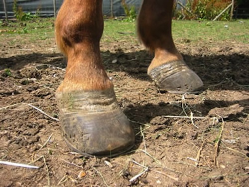
Dr Sue Dyson of the Animal Health Trust is Newmarket, England performs systematic lameness exams on horses referred to the clinic for diagnosis or treatment. (Sue Dyson/Hoofcare Publishing image)
Imagine Sue Dyson examining Thomas, a five-year-old warmblood, who competes in low-level showjumping.

Thomas was referred to the Animal Health Trust because he was reluctant to land from a fence with the right forelimb leading. He had become awkward turning to the left and sometimes felt "pottery" (unlevel or unstable) in front. Thomas's vet had detected some swelling in the region of the left front foot's coffin joint, just above the coronary band. He was sound on a straight line but on a circle he showed left forelimb lameness on the left rein, which was worse on a firm surface that a soft one.

Nerve blocks were used to desensitize the back of the foot and this eliminated the left forelimb lameness. On a separate occasion, an injection of a local anesthetic into the coffin joint also made Thomas sound.
The impar ligament of the navicular bone is known as the Distal Sesamoidean Impar Ligament or DSIL. It anchors the navicular bone by connecting it to the coffin bone (P3). This ligament is very deep inside the foot and is not visible on a radiograph. (HC Biovision plastination image, uesd with permission)
X-rays of the foot were taken, but the vet saw no abnormalities. The vet explained that although primary injuries of the coffin joint can occur, a positive response to nerve-blocking did not rule out the possibility of injury to one of the other related structures.

A treatment plan devised by the horse's regular vet directed that Thomas’s coffin joint was treated with an injection of corticosteroids and hyaluronan, which made him sound within a few days and able to resume full work. He continued to progress well for about six weeks but clinical signs recurred. Never blocks were repeated and yielded similar results.

This MRI shows what a normal, undamaged DSIL looks like. It appears like a solid bank of white leading from the navicular bone to the inside of the coffin bone. The veterinarian interpreting the MRI would hope to see the DSIL looking the one in this image. (Hallmarq reference image)
The vet examined the collateral ligaments of the coffin joint, just above the coronary band, and could see no abnormalities. It was therefore suggested that an MRI scan should be carried out to try to establish a more definitive diagnosis. MRI often identifies injuries which are invisible when using other techniques such as x-rays or ultrasound, particularly when the problem is in the foot.

The MRI scan, performed at the Animal Health Trust, revealed that Thomas had sustained an avulsion fracture—an injury to the bone in a place where a tendon or ligament attaches to it—at one side of the attachment of the distal sesamoidean impar ligament to the pedal (coffin) bone. Clearly this was a problem that was going to require rest before Thomas would happily swap leads again.

Damaged DSIL: Deciding to look into the foot in search of a root cause of the lameness was delayed for months with this horse, while the vet treated what might be the horse's problem. This type of injury is relatively rare but can be diagnosed through advanced imaging. The MRI showed that the foot was damaged in the region of the distal sesamoidean impar ligament (DSIL) in the navicular zone. Compare this reference image with the other images of the DSIL in this blog. (Image courtesy of Dr Martinelli, California Equine Orthopedics)

Thomas's case study is a sponsored blog post in cooperation with Hallmarq Veterinary Imaging.

Watch for more in the Hallmarq-sponsored article series on The Hoof Blog, and check their social media system and especially their info-deep web site for lots more information.

To learn more about Hallmarq Veterinary Imaging and standing MRI technology for horses:

• Become a fan  of the new Hallmarq Equine MRI Facebook page;
• Follow @HallmarqMRI on Twitter;
• Subscribe to the hallmarqvetimaging channel on YouTube.com;
• Watch for a growing equine distal limb Hallmarq MRI image gallery on Flickr.com;
• Visit the Hallmarq.net web site. (Plan to spend some time there!)


MRI images used in this article were provided by Hallmarq as examples and are not the actual radiographs from Thomas's file.

This case study originally was written for an article on lameness in Horse and Hound Magazine.


Follow the Hoof Blog on Twitter: @HoofcareJournal
Join the Hoofcare + Lameness Facebook Page

Saturday, June 19, 2010

CT Scans Added to Washington State University Vet School's Equine Imaging Options


Horse in CT Scanner
A Quarter horse mare recently underwent a spiral
CT scan to examine a mass near one of the
carotid arteries leading to her head
Washington State University's Veterinary Teaching Hospital (VTH) has installed a new spiral computed tomography (CT) scanner for use in both small and large animals, with funds donated by a generous family. 
The VTH has had a CT scanner for more than two decades and a magnetic resonance imaging machine (MRI) since 1996.  With the new Toshiba Aquilion 16-slice spiral CT unit operational, both MRI and CT in WSU's veterinary college are among the most advanced complementary tools for diagnostic imaging in the profession. 
 "Before the new CT, we only imaged a few horses a month  (using CT), but now I would expect to do 10 times that," said Professor John Mattoon, a board certified veterinary radiologist and chief of WSU's diagnostic imaging section.  "There were limitations with the old technology that hampered its everyday use, but the new CT is truly state-of-the-art, with brand new software that greatly improves its capabilities.  Our goal is to examine 100 horses a year with the CT, and several small animals a day."
Speed is one of the new CT's main features.  It can scan 1750 millimeters (mm) or about 5.75 feet of a patient's body in 1 mm slices in 38 seconds.  Twelve images per second are displayed and all 1750 digital images are delivered within three minutes.  The machine's resolution can "see" details as small as 0.35mm; a little more than 13/100ths of an inch.  The imaging is produced in a variety of planes as well as in three-dimensional representations of anatomic structures. A small animal can often be imaged in the new CT scanner in seconds, in many cases without general anesthesia.
"A horse with a complex fracture was examined with the new CT in early June and it was completed in a couple of minutes," Mattoon said.  "The anesthesia and prep-work it takes to get the horse into the machine takes much longer than the actual exam.  By comparison, MRI may take an hour or more. Still, these two imaging modalities are complementary to each other, and one does not necessarily exclude the use of the other."
Horses are too large to fit entirely in the CT scanner, so only the head, upper neck, and lower limbs are imaged.  For smaller animals, the entire body can be scanned, and is especially useful for examining the lungs and abdomen.
"CT scans are the first choice in human medicine for imaging the lungs and abdomen, and I think it should become the standard of abdominal imaging in smaller animals as well," said Mattoon, who has practiced radiology for more than 25 years.    
As a result of the CT's speed, animals have to spend much less time under anesthesia, if at all.  "For horses, we can use a short-acting anesthetic, and some small animals can just be sedated without undergoing anesthesia," Mattoon said.  "This is an important advancement because there are always risks associated with anesthetizing an animal.
"Overall, CTs at WSU should be less expensive because exams take less time and anesthesia.  This particular new CT scanner should also open up a whole new area of research, including vascular imaging and shunt studies. I imagine that in the beginning we will do a lot of cases in which we use both CT and MRI."
This brief video showcases Washington State's new CT service, as illustrated by the Quarter horse with the cartoid artery mass that needed to be imaged.
Information and elements for this article are the property of Washington State University.

19 June 2010 | Fran Jurga's Hoof Blog at Hoofcare.com
© Fran Jurga and Hoofcare Publishing

Please, no use without permission. Fran Jurga's Hoof Blog is a between-issues news service for subscribers to Hoofcare and Lameness Journal. This blog may be read online at the blog page, checked via RSS feed, or received via a digest-type email (requires signup in box at top right of blog page). To subscribe to Hoofcare and Lameness (the journal), please visit the main site, www.hoofcare.com, where many educational products and media related to equine lameness and hoof science can be found. Questions or problems with this blog? Send email to blog@hoofcare.com.