Showing posts with label preservation. Show all posts

Sunday, April 15, 2012

Trophy Hoof: Taxidermist Rowland Ward Preserved Part of a Favorite Horse Forever


Every once in a while, you'll see one in an antique store or on eBay: a preserved hoof from long ago. A favorite hunter or cavalry charger lives on because one of his hooves was preserved as a trophy.

The National Army Museum in London, England has their collection on display now as part of their "War Horse: Fact & Fiction" exhibition.

The two primary ones used in curator Pip Dodd's video lecture are excellent contrasts in hoof form and shoe fit.

Would you care for a cordial? Rowland Ward combined his big game expertise and his hoof trophy specialty in this mini-bar created from an African elephant's foot.


Now, here's my question: Did they only preserve one hoof? Would they have just tossed out the others? What if it was a really famous horse?

More recently, I've come to question the preparation of the hooves themselves. If British army regulations required hooves to be burned with identification numerals, and if the farriers were required to retrieve the numbered hooves after battle, why do we seldom see hooves with numbers burned into the wall? Were they sanded down until the numbers disappeared?

Were some preserved hooves merely vehicles to display horseshoes? This heart bar shoe is almost 100 years old; it lives in the beautiful tack room at the Badminton House stables in England. The hoof doesn't look particularly like it suffered badly from laminitis, although the beloved horse who wore the shoe apparently did. There may have been different priorities for hoof trophies.

Perhaps the answer lies here, in this blog's story from December 2011 about the dreaded farrier ax:

"The Household Cavalry still burns numbers into three out of four of each horse's hooves. The near hind bears the horse's army number, the near fore his squadron number and the off fore has the regiment's initials."

While some people are repulsed at the site of hoof trophies, others are intrigued. First of all, they were preserved in an era where a rider may not have had the option of a photo or even a drawing to remember a favorite horse.

But what intrigues Hoofcare + Lameness readers is that the final product of a trophy doesn't seem to have much--if anything--to do with how the horse was shod. The shoes attached to the trophies seem to have been crafted by silversmiths, not farriers. Their fit is questionable and some even lack nails, although the clinches can still be seen in the hoof wall.

Much more information on hoof preservation--not just of horse hooves but safari trophies and game--was detailed in the 1883 book, Observations on the Preservation of Hoofs and Designing of Hoof Trophies, by Rowland Ward of London and Nairobi.

Ward, who had aspired to become a sculptor in his youth, was quite a prolific trophy artist; he offered more than 50 designs for hooves in his shop. His designs were patented and his clients included the Duke of Edinburgh.

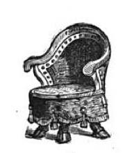
A Rowland Wolf hoof chair
He was a man of near and far ambitions; while his bread-and-butter might have been British horse hooves, his fame and fortune came from big game bagged by his wealthy clients--many of them Americans--on safari in Kenya.

The book details an important part of hoof trophy-making that has always challenged me. He states that the shoe worn by the horse is useless when making a trophy because the foot will change shape during the preservation process, and that crafting a suitable shoe for the trophy is part of the trophy-builder's task--and that a farrier is not the craftsman to be hired to build the shoe.

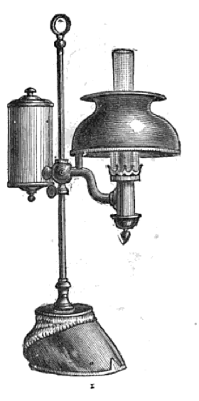
A Rowland Wolf hoof lamp
"The farrier's work should stop when the horse dies," he writes, "by which I mean that when the hoof is severed at the fetlock joint, for treatment by the naturalist, the farrier should not be allowed to take off the shoe, or in any other way to exercise his craft, either by cleaning of the flesh or skin or hoof, in any way whatever, and particularly not by boiling, or scalding or baking...."

He also mentions that part of the skill of the trophy-builder was in repairing the frogs of horses that had been affected by thrush, which apparently was prevalent in the hooves sent to him for preservation. Unfortunately, he doesn't go into much detail about how he did this.

Ward employed an assistant who worked on nothing but horse hoof trophies for more than 20 years.

Rowland Ward died in 1912 but the Rowland Ward business is still in business in Johannesburg, South Africa.  For some time, there was a US branch of the business, most recently headquartered in Dallas. The US office closed in 2009.

The company's web site is a mecca for what is left of the big-game hunters, and those who study the history of hunting and taxidermy, and the skillful arts of Rowland Ward.
A Rowland Wolf hoof scale

Today we have plastination and freeze-drying to preserve horse hooves, but the reason behind the preservation tends to be for educational purposes, rather than to preserve a memory or create a memorial. We demand lifelike detail, rather than artistic expression.

For Rowland Ward, a hoof from a dead horse was a blank canvas for artistic expression and his imagination ran as wild as the big game that arrived on his doorstep to be preserved, hooves and all.

To learn more:

National Army Museum (UK) hoof trophy feature

Sports Illustrated (1959): A Man Who Knows How To Stuff An Elephant

Farrier's Ax: A Museum Restores a Gruesome Tool of Mercy Designed to End the War for Horses 

Why Is That Guy Following Prince William and Kate Middleton Carrying a Big Shiny Ax? Because He's the Farrier, That's Why! 

 A collection of hoof trophies, including at least one by Rowland Ward, in the collection of the Canadian Anglo-Boer War Museum

© Fran Jurga and Hoofcare Publishing; Fran Jurga's Hoof Blog is a between-issues news service for subscribers to Hoofcare and Lameness Journal. Please, no use without permission. You only need to ask. This blog may be read online at the blog page, checked via RSS feed, or received via a digest-type email (requires signup in box at top right of blog page). To subscribe to Hoofcare and Lameness (the journal), please visit the main site, www.hoofcare.com, where many educational products and media related to equine lameness and hoof science can be found. 

Follow Hoofcare + Lameness on Twitter: @HoofcareJournal
Read this blog's headlines on the Hoofcare + Lameness Facebook Page
 
Disclosure of Material Connection: I have not received any direct compensation for writing this post. I have no material connection to the brands, products, or services that I have mentioned, other than Hoofcare Publishing. I am disclosing this in accordance with the Federal Trade Commission’s 16 CFR, Part 255: Guides Concerning the Use of Endorsements and Testimonials in Advertising.

Monday, October 12, 2009

Plastinate Anatomical Tools Make Everything Perfectly Clear

by Fran Jurga | 12 October 2009 | Fran Jurga's Hoof Blog


It's Columbus Day, so you're invited to "discover" a new equine anatomy reference tool that will be a boon to your ability to creatively and constructively communicate with colleagues, clients and students no matter where you are.

Plastination is a tissue preservation process that gained worldwide fame this decade with the Body Worlds museum exhibit. The last I heard, Body Worlds was set to overtake King Tut as the most viewed exhibit in the world. It shows human organs and muscles preserved in various positions or medical conditions. But everyone I know came out of it saying, "Wow, if they could just do that for horses..."

And someone has. Germany's veterinary anatomy expert Dr Christoph von Horst has patented a process for preserving veterinary specimen in this way; he's done birds and rats and ticks and dogs. But thanks to the encouragement of people like Dr. Chris Pollitt and a loud cheer from Hoofcare and Lameness, Dr Von Horst is preparing spectacular hoof and distal limb anatomy specimen, and you end up with a hoof music video slide show on a day of discovery.

I remember for years how I struggled trying to learn anatomy from textbooks. I couldn't get the 3-D part. I believe that 3-D models from HorseScience are the absolute way to learn and study anatomy and that they revolutionized my ability to understand the hoof, to the extent I can say that I do.

These plastination models are a step somewhere between anatomy models and an x-ray. They come in different models, designed for more or less portability. Many will slip inside a briefcase or agenda planner...or even a jacket pocket.

I can't wait for you all to see these teaching aids. They are like living x-rays...in equally-living color! They are actual paper-thin slices of tissue vacuum sealed inside layers of crystal clear acrylic resin.

You can keep one in your briefcase, or collect a set to show different conditions like laminitis, a navicular cyst, ringbone, etc. or use them to show where a shoe will sit, where you will trim (or won't trim) or where an injection or surgery site will access a joint or problem.

This specimen illustrates ringbone quite clearly but, like most anatomy models, the medical history of the animal is not available for reference. (Dr. Christoph von Horst image)

The plastinate tissue is very clear and well-defined because it is paper-thin and light passes through, illuminating the details and edges of structures and their relative textures. (Dr. Christoph von Horst image)

The specimen come in two types: flat sheets, which are about 3/8" thick, or the block versions, which are about 3/4" to 1" thick. The blocks are stunning and look fantastic on a desk or bookshelf, particularly if there's a light nearby. They make a beautiful gift or presentation award.

Of course, no two are alike. Hooves are available in sagittal, coronal and transverse sections, with the vast majority being sagittal, since that is the primary view people are accustomed to using for reference.

Right now we even have a foal's limb and a huge draft horse lower limb with what Dr Von Horst labels as lymphangitis-type swelling. There's also a stunning example of pastern ankylosis.

Even a large joint like the hock can be encapsulated into a plastinate specimen. (Dr. Christoph von Horst image)


Also available are laminated posters of several popular types of distal limb and hock plastinates; you can write on the plastic, draw a shoe or cast on, or use the poster for teaching by asking students to fill in labels for specific structures. Plastic casts of the blood supply and plaster casts of hooves are available by special order.

The best news? Prices start at under $100, plus shipping, with the blocks selling for about $200 at the current exchange rate.

Be sure to visit the Hoofcare and Lameness booth at conferences this fall to see these amazing teaching and learning aids, or contact the office to arrange an order to be selected and shipped directly to you.

If you have trouble with my video widget, you can also view the slide show on Hoofcare's slowly-expanding video channel. The widget seems to be skipping over some of the images in favor of text slides.

© Fran Jurga and Hoofcare Publishing. No use without permission. You only need to ask. Fran Jurga's Hoof Blog is a between-issues news service for subscribers to Hoofcare and Lameness Journal. This blog may be read online at the blog page, checked via RSS feed, or received via a digest-type email (requires signup in box at top right of blog page). To subscribe to Hoofcare and Lameness (the journal), please visit the main site, www.hoofcare.com, where many educational products and media related to equine lameness and hoof science can be found. Questions or problems with this blog? Send email to blog@hoofcare.com.