 |
 |
| Story protected by copyright. Thank you for sharing the link on social media and with your friends and colleagues. |
In spite of an all-time pinnacle of near-perfection in both ready made shoes and high tech materials to design and fabricate custom equine hoof orthotics for every imaginable injury or deformity, it is a foregone conclusion that 3D printing will play a role in the production of horseshoes in the future.
How do we make sure that the new technology heads in a direction that helps horses and makes better horseshoes, not just more horseshoes?
You can go online right now and download the code to 3D-print horseshoes, such as corrective shoes for foal limb deformities, but the output is pre-set to specific sizes and shapes.
The data driving 3D printing of horseshoes has, until this point, been largely two-dimensional, based on using Computer Assisted Drawing (CAD) software. The exception is some impressive strides made in the design and fabrication of prostheses for amputee horses, but that process is predicated by the requirement of optimum functionality in supporting the horse's weight and allowing movement, rather than trying to replicate a foot that no longer exists, or attach a shoe to it.
On the other end of things, we know we can also 3D print horse feet, using data from dead or living horse limbs. Professor Chris Pollitt in Australia can (and does) even "print" an entire distal limb model from CT scan data, using sophisticated modeling software. Commercial online 3D CAD websites offer printing plots for a coffin bone--but not a hoof--for downloading and printing.
 |
| Positioning of the foot is a critical stage early in the process. The software creates a detailed image of the hoof capsule. |
But the clinical applications for all this “available” technology is NQTY--Not Quite There Yet. Thesis after thesis from bioengineering scholars outlines the need and possible processes for scanning horse hooves in three dimensions so a shoe can be made to fit exactly. But the hoof-matching-shoe paradigm hasn’t been an integrated process.
In 2017, a research group in Poland published an Open Access, peer-reviewed article on a 3-D scanning method for comparison of the morphology of left and right limb coffin bones retrieved from the front feet of horses. They made no mention of the feasibility of their turntable-based system for scanning the feet of a living horse, even with a handheld scanner.
The closest thing to a clinical application was a thesis by Magdelana Rosendahl at Skovde University in Sweden in 2013. Her beautifully illustrated document is written in Swedish except for a brief English abstract, in which she predicts that the value of 3D scanning in the farriery profession will be its potential to professionalize a traditional trade or craft.
Rosendahl predicted the value in using 3D scanning would be to document cases over time, rather than as a clinical procedure for helping lame horses. Rosendahl believed that individual farriers would use 3D printing to provide evidence of the effects of their work and forecast a prognosis for ideal hoof balance and horse health in their clients' horses.
This year, we may have both the foot scan and the shoe design in one place for the same (living) horse, if trials in the forge of the Utrecht University vet school in The Netherlands produce the results expected.
In the latest example of the global trend for farriers to be directly involved in and even initiate equine research, Utrecht’s vet school resident farriers Jan de Zwaan and Gerben Bronkhorst are issuing an international invitation for fellow professionals to support their effort to create an integrated system that both scans the foot and designs a shoe to fit and support it as required by lameness, injury or conformation.
 |
| With the foot layer sent to the background, the shoe can be fine tuned on the screen and then sent to the printer. The farrier can see the shoe from any perspective. |
The team needs to raise 8,000 Euros ($8,993 USD) to complete the first stage of this research, which is outside the purview of any commercial interests for the end product.
The Hoof Blog was fortunate to do a question and answer session with farriers Jan de Zwaan and Gerben Bronkhorst of Utrecht University and project manager Dr. Inga Wolframm; their team also includes Professors Rene van Weeren and Harold Brommer.
 |
| For the purposes of demonstration, the team printed out both the foot and a sample shoe. |
Hoof Blog: Do you already have the software, scanner and 3D printer or is the purchase of the equipment part of the reason for the fundraising? I assume the software is a standard CAD-type application?
 |
A handheld
Artec scanner |
With regards to the funds we want to raise, we’re hoping to raise enough to cover the material costs for the printing and testing of a couple of additional prototypes of differently shaped shoes, such as a straight bar shoe, heart bar shoe, etc., to test the ‘proof of principle’ and subsequently to print and test six pairs of shoes, to be used initially on our own horses. (These are horses owned by the faculty, and are ridden and trained by our veterinary students.)
 |
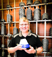
Gerben Bronkhorst, one of two
farriers at Utrecht University
vet school.
|
At this point, we want to focus on gathering as much data as possible in order to come up with a 3D printed shoe that really improves equine/equid welfare. Once we’ve done that, we’ll think about the next step. Most importantly, though: As a university, we feel that it’s our responsibility to develop new, innovative ideas for the benefit of society (or parts of society).
 |
The challenging weak foot of the barefoot horse
described in the video.
|
UU: Jan and Gerben think that a cuff is not only necessary but beneficial. That’s because the cuff can be printed to take on the exact shape of the hoof wall, thus allowing us to fit the shoe (and the cuff) to the millimeter. We think that this will also ensure that the glue will adhere for longer (as the more snug the cuff, the less likely it’ll be to let go of the hoof). The idea is to also develop a shoe that allows for a farrier to either use glue or nails, or both.
HB: Readers might think you are suggesting making a mold of the foot from scanning. I believe the foot scans will live in the software, and the mold you show in the video is to illustrate that the shoe fits the foot for the video. The shoe is the product, not a mold of the foot. Is that correct?
UU: Yes, that’s precisely what it is. The mold you see in the video was indeed made to help us in the development of the prototype. In the project we’ve got in mind now though, we’ll only have to scan the foot, save it to the software program, and print from there.
HB: What is the approximate time required from scanning the foot to having a shoe in hand, ready to be glued on? (Once a user develops proficiency)
UU: At this point, it’ll take approximately 12 hours. The scanning only takes a couple of minutes, the design process of the shoe in the software (so the adding of the various features, depending on the biomechanical/orthopedic requirements) takes around an hour, but might become easier with more proficiency. The printing itself takes nine hours, and that’s the “delay”, if you can call it that. Obviously, the development doesn’t stand still here either, so faster printers will result in faster shoes.
 |
| Utrecht University vet school farrier Jan de Zwaan wants the end product printed horseshoe to have options be either nailed or glued, according to the needs of the horse being treated in the clinic. |
Jan de Zwaan also commented that the output of a shoe predetermined to be glued rather than nailed to the foot was not a prerequisite; they may nail or glue 3D-printed shoes on a case by case basis.
Once the computer-designed shoe is applied to the horse’s foot, its effect on the horse will be measured objectively. Jan and Gerben will use the university’s state-of-the-art movement analysis systems to monitor the horses' stance and movement patterns prior to and following the shoeing.
• • • • •
If you would like to be part of this project, the Utrecht team would love to receive your donation.
 |
| Click here to access the project page. |
Alternately, the university offers a crowdsourcing form of fundraising, which allows donors to start a team of sub-donors, or an individual donor can add his or her donation to a pre-existing group’s fundraising campaign.
The project has raised 11% of its goal, at the time this article was written.
Click here to visit the fundraising page to see the project's progress and add your donation, of any amount. US donors may need to use the PayPal link, which will work with any credit card. All donors who supply contact information will be kept informed of progress as the project continues.
If you have questions about funding for this project, please email Dr. Inga Wolframm, Head of Fundraising for the Faculty of Veterinary Medicine at Utrecht University: i.wolframm@uu.nl.
3D Horseshoes research project information page on the Utrecht University website
3D Horseshoes research project donation page on the Utrecht University website
3-D Printing in the Forge and Clinic: Hoof Anatomy models, Veterinary Applications, and Horseshoes (Hoof Blog archives)
Short informal talk by MIT sports technology grad Dr. Mike Vasquez on his experience applying 3D printing in sports
Laminitic Pony in Australia First Horse in History to Wear 3D Printed Titanium Horseshoes (Hoof Blog archives)
Chris Pollitt's Laminitis Images Have a New Look: MIMICS Software Goes 3-D (Hoof Blog archives)
Paśko, S., Dzierzęcka, M., Purzyc, H., Charuta, A., Barszcz, K., Bartyzel, B. J., & Komosa, M. (2017). The Osteometry of Equine Third Phalanx by the Use of Three-Dimensional Scanning: New Measurement Possibilities Scanning, 2017.
Rosendahl, Magdelena. (2013). Underlätta bearbetning av hästskor. (Facilitate the machining of horseshoes.) (Hoof Scanner) Thesis, Skovde University, Sweden.
Additional articles:
Preece, D., Williams, S. B., Lam, R., & Weller, R. (2013). “Let's get physical”: advantages of a physical model over 3D computer models and textbooks in learning imaging anatomy. Anatomical sciences education, 6(4), 216-224.
Thomas, D. B., Hiscox, J. D., Dixon, B. J., & Potgieter, J. (2016). 3D scanning and printing skeletal tissues for anatomy education. Journal of anatomy, 229(3), 473-481.
• • • • •
 |
| If requesting via email, be sure to mention that you want your name added to the email list. The email subscription for The Hoof Blog is free of charge. |
© Fran Jurga and Hoofcare Publishing; Fran Jurga's Hoof Blog is the news service for Hoofcare and Lameness Publishing. Please, no re-use of text or images on other sites or social media without permission--please link instead. (Please ask if you need help.) The Hoof Blog may be read online at the blog page, checked via RSS feed, or received via a headlines-link email (requires signup in box at top right of blog page). Use the little envelope symbol below to email this article to others. The "translator" tool in the right sidebar will convert this article (roughly) to the language of your choice. To share this article on Facebook and other social media, click on the small symbols below the labels. Be sure to "like" the Hoofcare and Lameness Facebook page and click on "get notifications" under the page's "like" button to keep up with the hoof news on Facebook. Questions or problems with the Hoof Blog? Click here to send an email hoofblog@gmail.com.
Follow Hoofcare + Lameness on Twitter: @HoofBlog
Read this blog's headlines on the Hoofcare + Lameness Facebook Page
Disclosure of Material Connection: The Hoof Blog (Hoofcare Publishing) has not received any direct compensation for writing this post. Hoofcare Publishing has no material connection to the brands, products, or services mentioned, other than products and services of Hoofcare Publishing. I am disclosing this in accordance with the Federal Trade Commission’s 16 CFR, Part 255: Guides Concerning the Use of Endorsements and Testimonials in Advertising.


